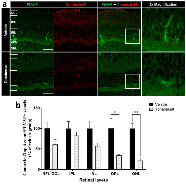
Figure 4.
(a) Immunohistochemical images showing an example of PLVAP (green) and connexin43 (red) co-labelling. (b) Tonabersat treatment decreased the number of connexin43 spots on PLVAP+ vessels within the OPL and ONL but not the NFL-GCL, IPL, and INL. Data are presented as a mean + SEM. Statistical analyses were carried out using two-way ANOVA with Sidak’s multiple comparison’s test. * p ≤ 0.05, ** p ≤ 0.01. Scale bar = 50 µm. n = 8–12 eyes per group.

