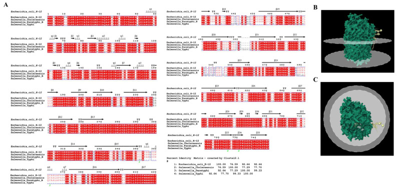
Figure 5.
FhuA receptor sequence analysis and protein structure view. (A) Amino acid sequences were aligned to identify conserved outer membrane residues among fp01 host strains. The comparison was performed for the FhuA receptor from E. coli K-12 (reference), S. enterica Choleraesuis, Typhi, and Paratyphi B serovars. (B,C) E. coli K-12 FhuA receptor 3D structure was obtained from Protein Data Bank (PDB).

