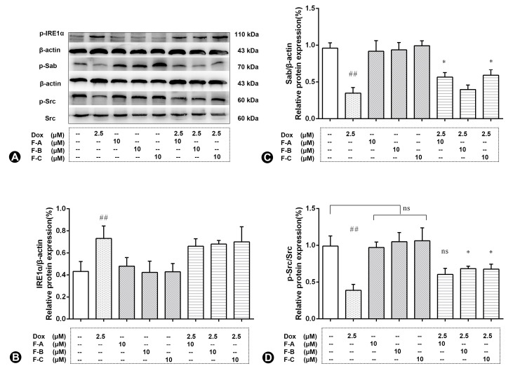
Figure 8.
The effects of three flavonoids on Sab, p-Src, and p-IRE1α protein expression level of Dox-treated H9c2 cells. Cells were pretreated with same concentrations of F-A, F-B, and F-C (10 µM) for 1 h, then exposed to Dox for 24 h. Relative folds of Sab (A,B), p-Src (A,D), and p-IRE1α (A,C). (B) The relative expression level of p-IREα in Dox group significantly increased compared to the NC group (p < 0.01). There was no significant activation of p-IREα in the F-A, F-B, and F-C group without Dox. F-A, F-B, and F-C had no significantly protective effect to reduce Dox-induced p-IREα protein phosphorylation. (C) The relative expression level of Sab in Dox group significantly decreased compared to the NC group (p < 0.01). There was no significant change of Sab in the F-A, F-B, and F-C group without Dox. But F-A and F-C can significantly enhance the Dox-induced reduction of Sab in comparison to the Dox model group (p < 0.05). (D) The relative expression level of p-Src in Dox group significantly decreased compared to the NC group (p < 0.01). There was no significant change of p-Src in the F-A, F-B, and F-C group without Dox. But F-B and F-C can significantly enhance the Dox-induced reduction of p-Src in comparison to the Dox model group (p < 0.05). Results are mean ± SEM from three independent experiments. ## p < 0.01 vs. control, * p < 0.05 vs. Dox group. ns, not significant.

