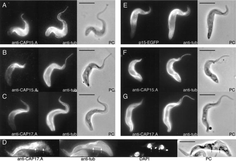
Figure 3.
Immunofluorescence analysis of CAP15/CAP17 expression showing labelling localized to the anterior portion of the cells. T. brucei bloodstream forms (A) or procyclic forms (B-D), and T. brucei procyclic forms overexpressing recombinant EGFP-tagged CAP15 (E), CAP15 (F), or CAP17 (G) proteins were labeled with anti-CAP15.A (A, B, and F) or anti-CAP17.A antisera (C, D, and G) and a monoclonal anti-tubulin TAT1 (A-G). The recombinant GFP-tagged CAP15 (E) was visualized by fluorescence analysis. In panel D, the cells were also stained with DAPI, and both extremities of the mitotic spindles are showed by arrows. The cell in panel D shows that, although the subpeculiar, flagellum, and spindle microtubules are labelled by anti-Cap17.NA antisera, the spindle is not labelled by anti-CAP17. Respective phase contrast (PC) images are also shown. Bar, 5 μm.

