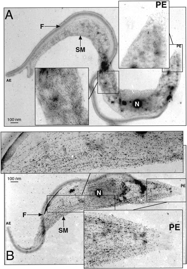
Figure 5.
CAP15/17 are associated with the subpellicular corset. Electron microscope analysis of the wild-type (A) and CAP17 overexpressing (B) T. brucei procyclic forms with the anti-CAP17 immune serum. Insets represent a higher magnification (× 2.5) of the region with low (posterior end) or high levels of CAP17 in the wild-type cells, and the same regions in the CAP17 overexpressing cells, which show a high level of CAP17 labelling . The insets also show the microtubule labelling pattern of the gold particles. The gold particles clearly bind to microtubules and follow the helical path of the subpellicular microtubules along the length of the cytoskeleton. In wild-type (A) cells, the labelling is on the major portion of the cytoskeletal microtubules but show no, or extremely low, labelling at the posterior end. In CAP17, overexpressing (B) cells immunolabelling is on the whole of the subpellicular microtubules. AE, anterior end; PE, posterior end; F, flagellum; N, nucleus; SM, subpellicular microtubules.

