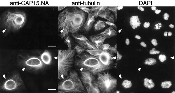
Figure 6.
Immunofluorescence analysis of CHO-K1 cells expressing CAP15. CHO-K1 Tet-ON cells transfected with the pTRE2-CAP15 plasmid and induced with doxycycline were stained with anti-CAP15.NA serum, monoclonal anti-tubulin, and DAPI. This shows that CHO-K1 cells expressing CAP15 (cells labelled with white arrowheads) have bundles around the nucleus, whereas no perinuclear bundles are observed for those not expressing CAP15 (unlabelled cells). Bar, 10 μm.

