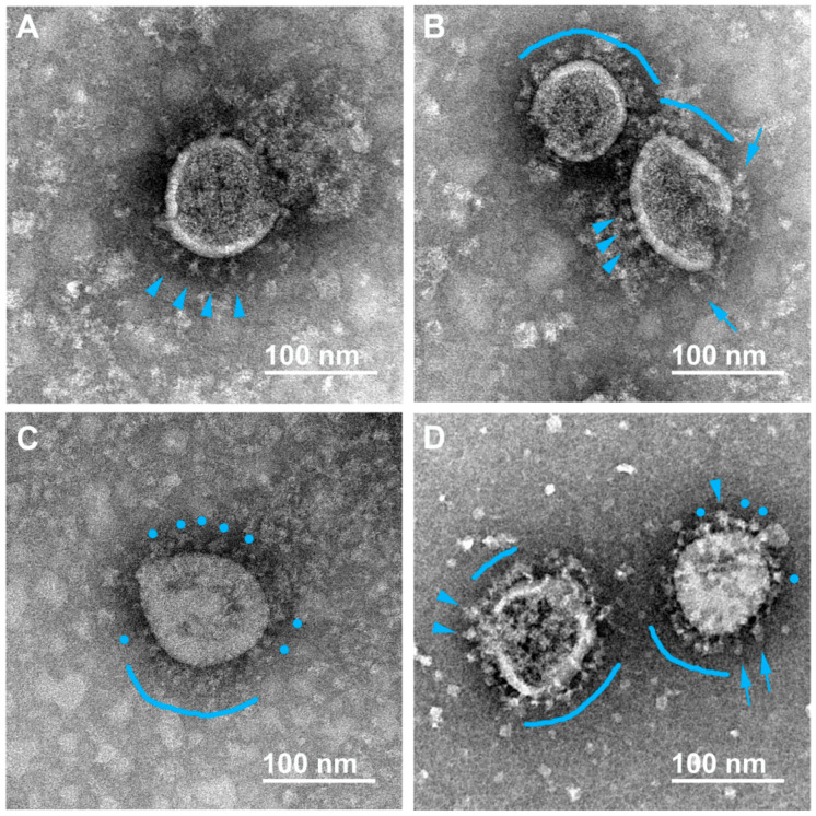
Figure 1.
Electron microscopy images of SARS-CoV-2 virions inactivated with 2% formaldehyde (1 h, 37 °C). Various forms of flail-like spikes supposedly corresponding to pre-fusion conformation are observed (A–D); single “classical” flails (A,B,D) and distorted/rounded flails (C,D) are indicated with blue arrowheads and blue circles, respectively. The flail-like spikes essentially tilted to the viral membrane are indicated with blue arrows (B,D). Coats of flail-like spikes are marked with blue arches (B–D).

