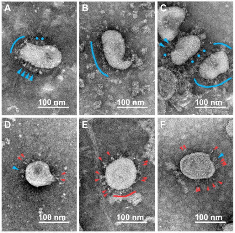
Figure 2.
Electron microscopy images of SARS-CoV-2 virions subjected to inactivation with BPL (1:2000, 36 h, 4–8 °C). Single spikes of different morphologies are distinguishable on the virions’ surfaces. Flails (blue arrowheads), rounded flails (blue circles), and flail coats (blue arches) supposedly correspond to the pre-fusion conformation of S-trimers (A–C), while needles (red arrowheads) and their coats (red arches) supposedly correspond to the post-fusion conformation of S-trimers (D–F).

