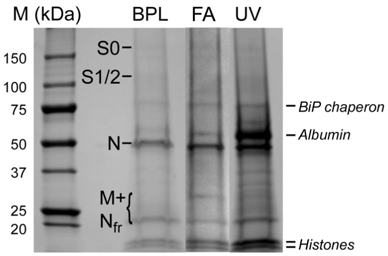
Figure 4.
Electrophoregram of preparations of viral particles inactivated by BPL (1:2000 for 36 h), formaldehyde (FA, 2%), or UV-radiation (UV, 3 min). Prior to loading onto the gel, the samples were concentrated via ultracentrifugation through a 20% sucrose cushion. Shown are the positions of SARS-CoV-2 structural proteins: S0—the non-split precursor form of S; S1/S2—cleavage products of S; M—membrane protein; N—nucleoprotein; Nfr—nucleoprotein’s fragments. Some admixed proteins are also indicated: BiP chaperon, albumin, and histones. All proteins were identified using Mascot searches after in-gel trypsin hydrolysis of protein bands, elution of peptides, and their MALDI-TOF-MS analysis.

