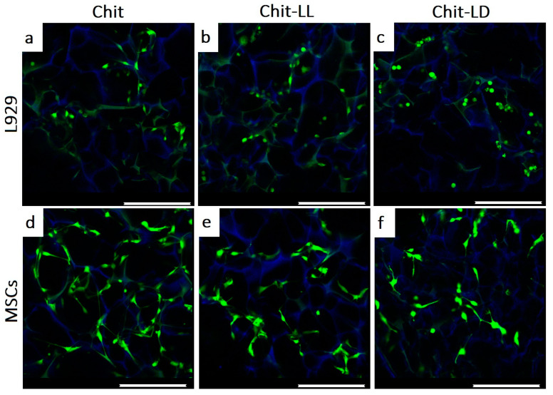
Figure 7.
CLSM images of the L929 mouse fibroblasts (a–c) and MSCs (d–f) after cultivation in the macroporous hydrogels from chitosan (Chit), chitosan-g-oligo(L,L-lactide) (Chit-LL), and chitosan-g-oligo(L,D-lactide) (Chit-LD) hydrogels for 3 days. The alive cells (in green) and the hydrogels (in blue) were stained with calcein AM and DAPI, respectively. The scale bar is 250 µm.

