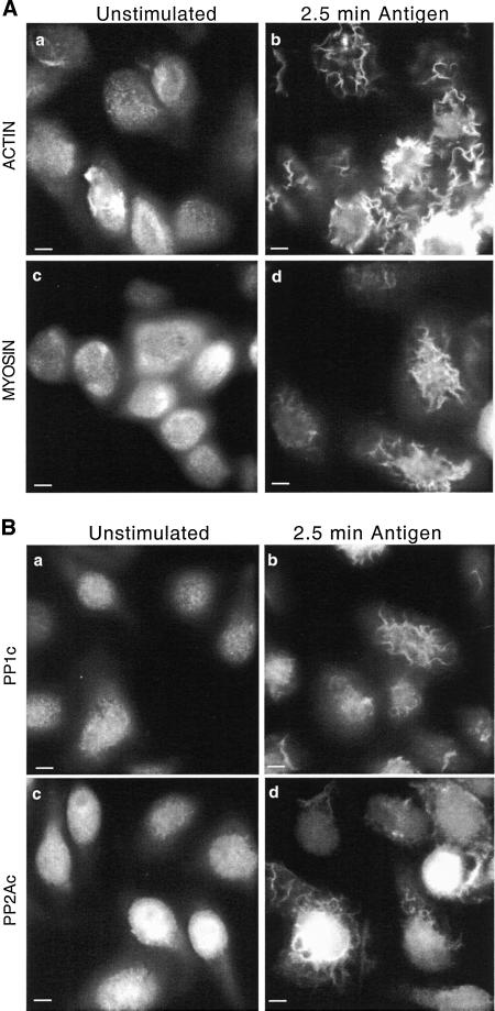
Figure 1.
Redistribution of actin, myosin, PP1, and PP2A to apical ruffles after antigen stimulation. Adherent monolayers of RBL-2H3 cells that had been incubated overnight with DNP-specific IgE were incubated in buffer alone for 2.5 min (unstimulated, a and c) or with antigen (100 ng/ml DNP-BSA) for 2.5 min (b and d), fixed, and proteins on the apical surface detected as detailed in MATERIALS AND METHODS. Actin (A, a and b) was detected using phalloidin-FITC and myosin (A, c and d), PP1c (B, a and b), and PP2Ac (B, c and d) by using specific polyclonal antibodies and FITC-labeled secondary antibodies. The images focus on the apical surface of the cells and are representative of those seen in at least five separate experiments. Bar, 5 μm.

