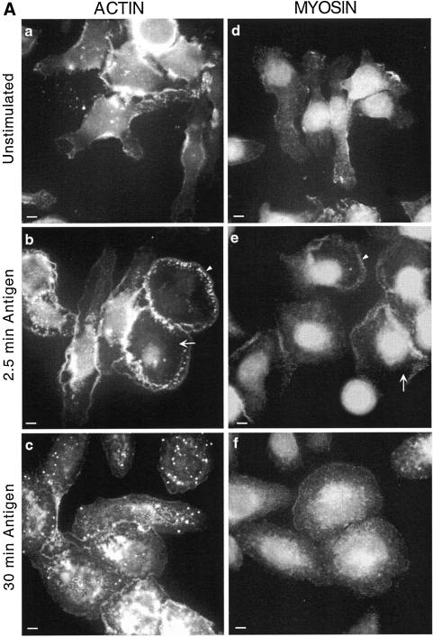
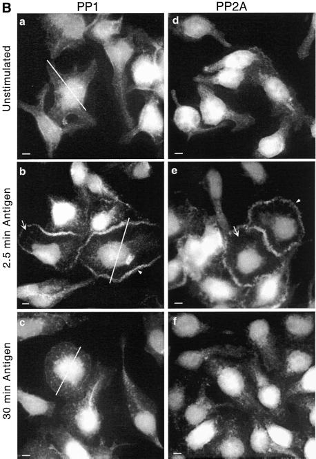
Figure 2.
Transient redistribution of actin, myosin, PP1, and PP2A to the periphery of the spreading cell after antigen stimulation. Adherent monolayers of RBL-2H3 cells that had been incubated overnight with DNP-specific IgE were incubated in buffer alone for 2.5 min (unstimulated, a and d), or with antigen (100 ng/ml DNP-BSA) for 2.5 min (b and e) or 30 min (c and f). The cells were then fixed and proteins on the basal cell surface detected as detailed in MATERIALS AND METHODS. Actin (A, a–c) was detected using phalloidin-FITC and myosin (A, d–f), PP1c (B, a–c) and PP2Ac (B, d–f) with specific polyclonal antibodies and FITC-labeled secondary antibodies. Arrows highlight regions where protein has cleared from the cytoplasm and arrowheads highlight the band of protein around the peripheral edge. The lines across the cells immunostained with PP1c were used to generate the fluorescence intensity graphs of Figure 3A. The images focus on the basal surface of the cells and are representative of those seen in at least five separate experiments. Bar, 5 μm.


