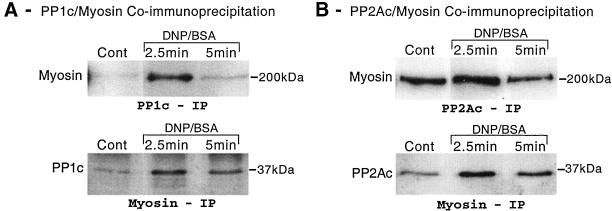
Figure 5.
Coimmunoprecipitation of myosin and PP1/PP2A after antigen stimulation. Adherent monolayers of RBL-2H3 cells that had been incubated overnight with DNP-specific IgE were incubated in buffer alone or activated with antigen (100 ng/ml DNP-BSA) for 2.5 or 5 min. Cells were lysed and PP1c, PP2Ac, or myosin immunoprecipitated using specific antibodies as detailed in MATERIALS AND METHODS. PP1c and PP2Ac immunoprecipitates were probed for myosin by Western blotting and myosin immunoprecipitates were probed for PP1c and PP2Ac as described above. The images are representative of those seen in two to three separate experiments.

