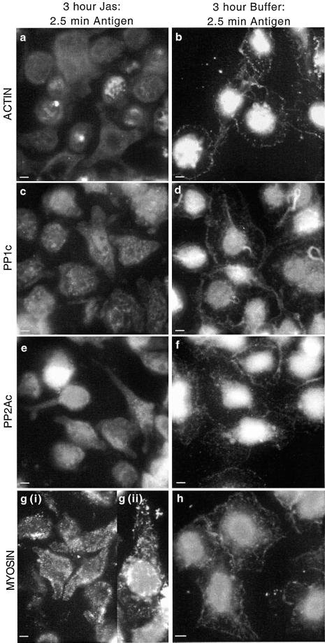
Figure 7.
Effect of jasplakinolide on relocation of actin, PP1, PP2A, and myosin after antigen stimulation. Adherent monolayers of RBL-2H3 cells that had been incubated overnight with DNP-specific IgE were incubated in a buffer containing 5 μM jasplakinolide for 3 h and activated with antigen (100 ng/ml DNP-BSA) for 2.5 min (a, c, e, and g). A separate set of cells was left for 3 h in buffer alone as a control and activated with antigen (100 ng/ml DNP-BSA) for 2.5 min (b, d, f, and h). All cells were fixed and proteins were detected as detailed in MATERIALS AND METHODS. Actin (a and b) was detected using an anti-actin mAb and FITC-labeled secondary antibody; PP1c (c and d), PP2Ac (e and f), and myosin (g and h) were detected using specific polyclonal antibodies and FITC-labeled secondary antibodies. Panel g (ii) is an enlarged view of jasplakinolide-treated unstimulated cells highlighting the short rods of myosin. The images focus on the basal surface of the cells and are representative of those seen in at least three separate experiments. Bar, 5 μm.

