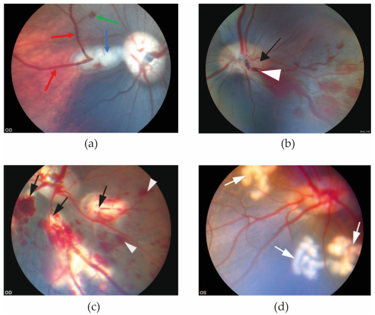
Figure 1.
Funduscopic images obtained 20 min after RVO was induced. (a) Eye with experimental BRVO. Retinal hemorrhage (green arrow) and dilation of retinal veins (red arrows) are observed upstream of the site of the occlusion (blue arrow) [25]. (b) Experimental CRVO can be created by application of laser around a retinal vein at the border of the optic nerve head (black arrow) followed by application of laser directly on the vein, displacing thrombotic material towards the lamina cribrosa. (c) Experimental CRVO can also be created by complete occlusion of 3–4 branch retinal veins, creating a condition in which the entire retina is affected by occlusion. Complete occlusion of 3–4 branch retinal veins results in severe ischemia constituting a severe ischemic CRVO. Laser-induced occlusion (black arrows) resulted in dilation of the occluded veins upstream of the occlusion sites. Flame-shaped hemorrhages (white arrowheads) developed shortly after the vein occlusions were induced [32]. (d) Control eye. Control eyes can be created by generating areas of laser applications similar to the RVO eye, using the same amount of energy and number of applications as in the RVO eye (white arrows), but without inducing occlusion. This control eye serves as control eye for (c) [32].

