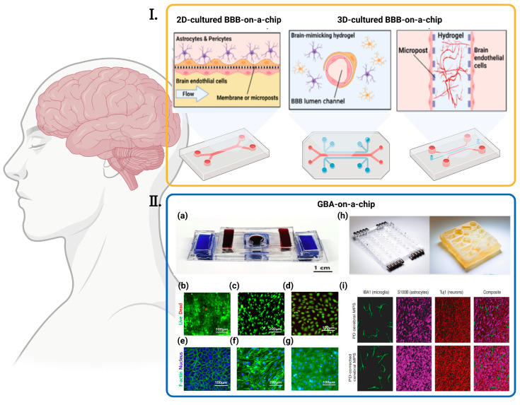
Figure 5.
Various BBBs-on-a-chip and GBA-on-chip. (I) Strategies for reconstructing the in vitro BBB in an OOC platform are diverse. Cells can be cultured in a 2D environment under fluidic flow or 3D-cultured with different approaches, such as seeding in hollow hydrogel or the angio/vasculogenesis approach [27] ©Copyright 2020, MDPI. (II) Various GBA-on-chip. (a) The picture of assembled GBA chip [30] ©Copyright 2020, Elsevier; (b–g) Fluorescent images of cells seeded in the chip [30] ©Copyright 2020, Elsevier: (b) Live/Dead Images of Caco-2, (c) bEnd.3, and (d) hBMECs when co-cultured (green = live, red = dead). F-actin/nucleus stain images of (e) Caco-2 cells, (f) bEnd.3 cells and (g) hBMECs when co-cultured (blue = nucleus, green = F-actin); (h) Left: pneumatic plates machined in acrylic; Right: mesofluidic plate machined from monolithic polysulfone [82] ©Copyright 2021, Amer Assoc Advancement Science; (i) Representative, 3D rendered confocal images of the PD (top) and control PD-C (bottom) cerebral MPSs composed of hiPSC-derived microglia (green), astrocytes (purple), and neurons (red) cocultured on 0.4-μm microporous 24-well Transwells [82] ©Copyright 2021, Amer Assoc Advancement Science.

