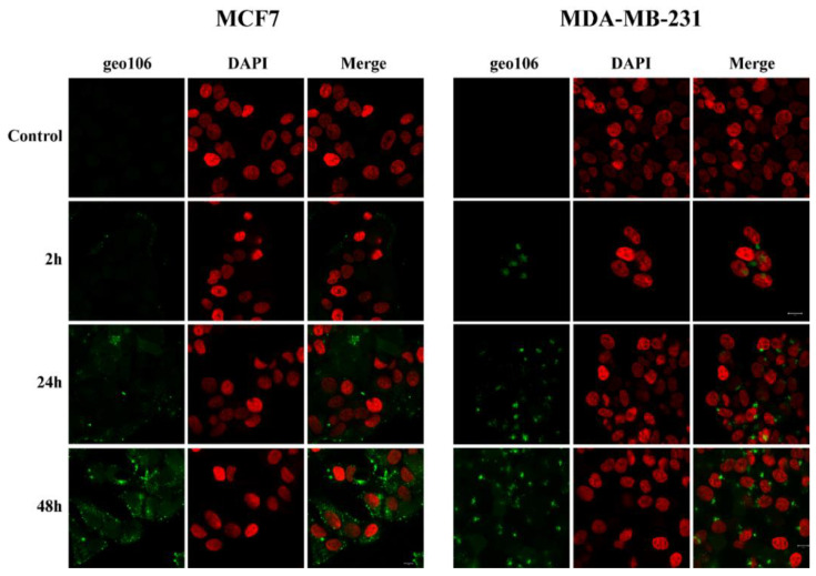
Figure 7.
Confocal microscopy images of MCF7 and MDA-MB-231 cells after incubation with 5 μM of geo106 for 2, 24 and 48 h, respectively. Cells were fixed with 4% paraformaldehyde. DNA was stained with DAPI (pseudo color red). Cells were visualized with a Zeiss LSM 780 confocal microscope using the Zen 2011 software.

