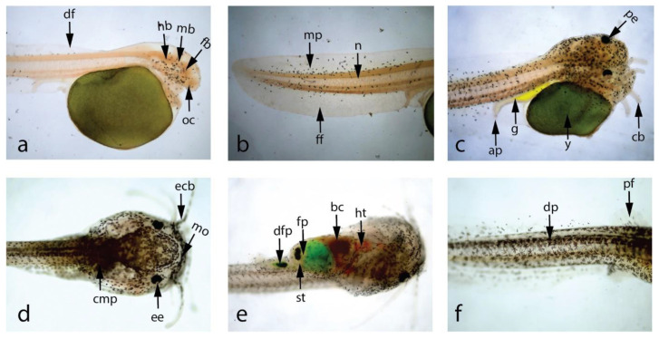
Figure 3.
Photomicrographs showing larval development of stinging catfish, (40×). (a) Newly hatched larva with oval-shaped yolk sac, (b) Posterior end of newly hatched larva, (c) 36 h larva with pigmented eye, (d) Dorsal view of 9 d larva, (e) Ventral view of 9 d larva, (f) Dense pigmentation on 11 d larva body, (ap—anal pore, cb—chin barbels, cmp—concentrated melanophores, df—dorsal fin, dfp—digested food particles, dp—dense pigmentation, ecb—elongated chin barbells, ee—epicanthus of eye, fb—fore brain, ff- fin fold, fp—food particles, g—gut, hb—hind brain, ht—heart, mb—mid brain, mo—mouth, mp—melanophores, oc—optic cup, pe—pigmented eye, pf—pectoral fin bud, st—stomach, y—yolk sac.

