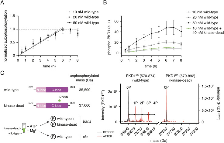
Fig. 3.
PKD1 activation loop autophosphorylation occurs in cis. (A) Activation loop autophosphorylation kinetics of PKD1KD at low concentration. Error bars are the SD of three biologically independent experiments. (B) Activation loop autophosphorylation of kinase-dead PKD1KD D706N in the presence and absence of excess wild-type PKD1KD. Error bars are the SD of three biologically independent experiments. (C) Mass spectrometry analysis of the autophosphorylation reaction containing 10 nM wild-type PKD1KD and 40 nM kinase-dead PKD1KD D706N shown in panel (B) Phosphospecies of wild-type PKD1KD are separated by 80 Da (double-headed arrows).

