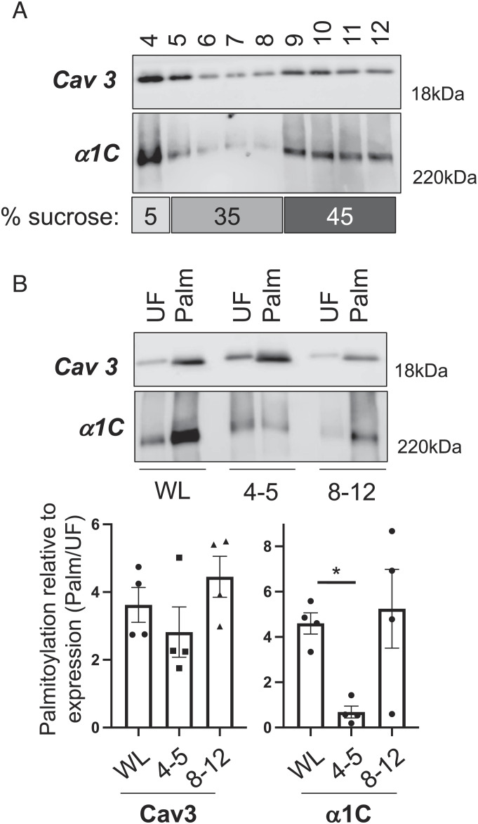
Fig. 2.
Palmitoylation and subcellular localization of the Ca(v)1.2 α1C subunit. (A) Western blot of caveolae prepared from mouse ventricular myocytes using a standard discontinuous sucrose gradient (sucrose concentrations in the numbered gradient fractions are indicated under the blots). Caveolin-enriched membranes were harvested from gradient fractions 4 and 5. (B) Palmitoylated proteins were prepared from whole cardiac lysates (WL) and pooled buoyant caveolar (4+5) and dense noncaveolar (8−12) fractions. The bar charts show the amount of Ca(v)1.2 α1C and caveolin 3 (Cav3) in the purified palmitoylated fraction (Palm) relative to the corresponding unfractionated whole lysate/pooled gradient fractions (UF). Caveolin 3 is equally palmitoylated in all fractions, but little Ca(v)1.2 α1C present in buoyant caveolar membranes is less palmitoylated. *P < 0.05 compared to WL, ANOVA followed by Dunnett’s multiple comparisons test, N = 4.

