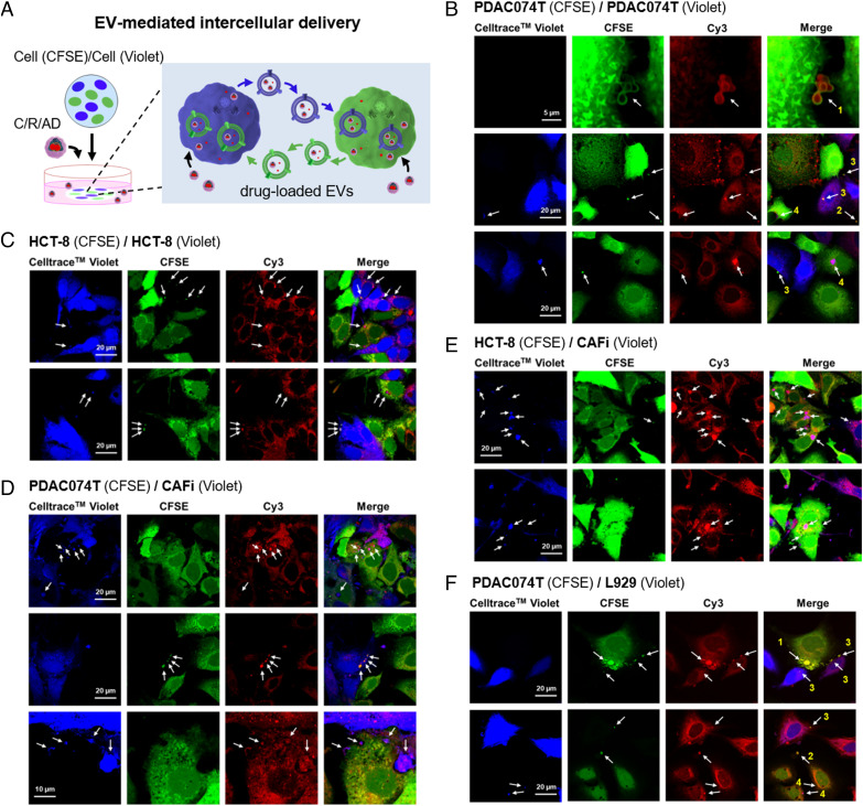
Fig. 4.
Intercellular Delivery of Cargo-Loaded EVs. (A) Cartoon illustration of the intercellular delivery of cargo-loaded EVs studied using confocal imaging. Cells were prestained with either CFSE (green) or CellTrace™ Violet (blue) to allow easy distinction in confocal imaging. (B–F) Confocal imaging revealed that Cy3-loaded EVs were transported between cells in the co-culture of PDAC074T (CFSE, green) / PDAC074T (CellTrace™ Violet, blue) (B), HCT-8 (CFSE, green) / HCT-8 (CellTrace™ Violet, blue) (C), PDAC074T (CFSE, green) / CAFi (CellTrace™ Violet, blue) (D), HCT-8 (CFSE, green) / CAFi (CellTraceTMViolet, blue) (E) and PDAC074T (CFSE, green) / L929 (CellTrace™ Violet, blue) (F), after incubation with C/R/AD. CAFi and L929 denote cancer-associated fibroblast and normal fibroblast, respectively. Arrows point to the Cy3-loaded EVs and show for example in B and F, arrows 1, the EVs (green) released by the green PDAC074T cells (CFSE) with Cy3-fluorescence (red) localized in their interior; arrows 2, the release of EVs (green) bearing Cy3 fluorescence from the green PDAC074T cells (CFSE) into the intercellular space; arrows 3, the Cy3-loaded EVs (green) released by the green PDAC074T cells (CFSE) and taken up by the blue PDAC074T cells (CellTrace™ Violet); arrows 4, the Cy3-loaded EVs (blue) released by the blue PDAC074T cells (CellTrace™ Violet) and taken up by the green PDAC074T cells (CFSE).

