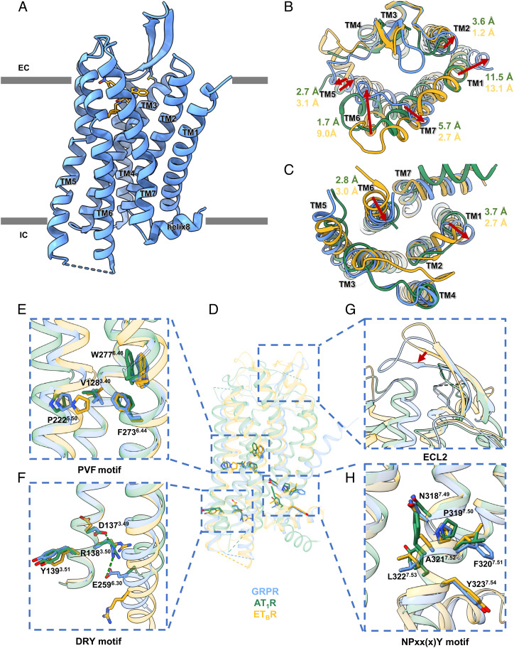
Fig. 1.
(A) Overall structure of GRPR-PD176252, with GRPR in blue cartoons and PD176252 in orange sticks. (B–H) Structural comparisons among GRPR (in blue), AT1R (in green), and ETBR (in goldenrod) from the extracellular view (B) and the intracellular view (C), with enlarged views of the conserved P5.50V(I)3.40F6.44 motif (E), D3.49R3.50Y3.51 motif (F), ECL2 (G), and N7.49P7.50xx(x)Y7.54 motif (H).

