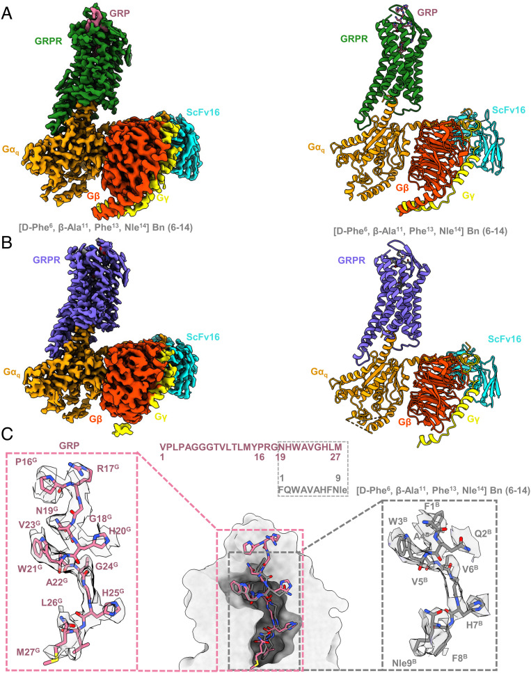
Fig. 3.
Cryo-EM structure determination of GRPR-Gq signaling complex. (A and B) Orthogonal views of the maps (Left) and models (Right) for the GRP (pale violet red) or [D-Phe6, β-Ala11, Phe13, Nle14] Bn (6–14) (dark grey) bound GRPR-Gq complexes, with GRPR was colored in forest green and medium slate blue, respectively. Gαq, Gβ, Gγ, and ScFv16 were colored in orange, orange red, yellow, and cyan, respectively. (C) Binding poses and sequence alignment of GRP and [D-Phe6, β-Ala11, Phe13, Nle14] Bn (6–14). The density maps of ligands were highlighted.

