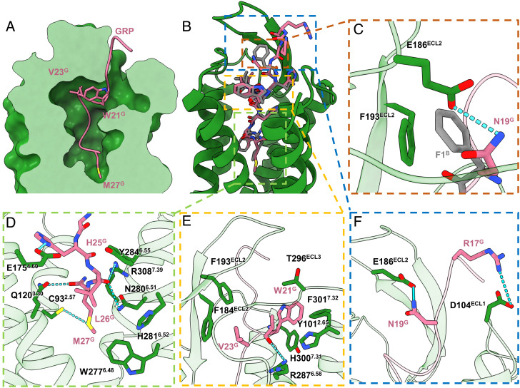
Fig. 4.
Peptide agonists binding to GRPR. (A) Cross-section of the peptide-binding pocket of GRPR was shown as forest green surface, with GRP as pink ribbons and side chains of W21G, V23G, and M27G displayed as pink sticks. (B and C) Structural alignment of GRP (green) and [D-Phe6, β-Ala11, Phe13, Nle14] Bn (6–14) (gray), with enlarged views of detailed interactions at N19G and F1B.(D) The TM bottom pocket. (E) The hydrophobic cavities around the helical fragment of GRP. (F) The extracellular loops.

