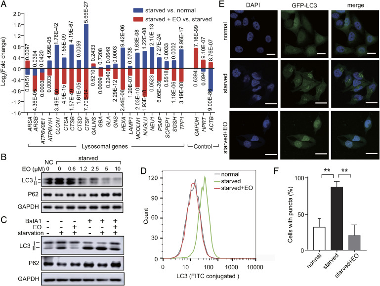
Fig. 5.
EO blocks the starvation-induced autophagy in a dose-dependent manner. (A) Expression analysis of lysosomal genes in HeLa cells treated with normal medium, nutrient-deprived EBSS medium with or without EO (n = 3, N = 1) according to RNA-Seq. Housekeeping genes GAPDHHPRT, and ACTB were used as controls. P values were marked accordingly. (B) Immunoblot analysis of LC3-I, LC3-II, and SQSTM1 (p62) in HeLa cells treated with normal medium control (NC), or nutrient-deprived EBSS medium with gradient EO (0 to 10 µM). (C) Immunoblot analysis of LC3-I, LC3-II and SQSTM1 (p62) in HeLa cells treated with normal medium, nutrient-deprived EBSS medium with or without EO (10 µM). BafA1 was used at a concentration of 200 nM. For B and C, GAPDH served as a loading control. One representative result of three independent experiments is shown. (D) Flow cytometric analysis of LC3 in HeLa cells treated with normal medium, nutrient-deprived EBSS medium with or without EO (10 µM). Using fluorescein isothiocyanate (FITC) conjugated LC3 antibody, the formation of lipidated LC3-II was quantified by flow cytometry. (E) Representative confocal images of HeLa cells stably expressing GFP-LC3-(ATG4 cleavage site)-RFP treated with normal medium, nutrient-deprived EBSS medium with or without 10 µM EO. Nuclei were stained using 4′,6-diamidino-2-phenylindole (DAPI). (Scale bars, 20 µm.) (F) Quantification of the percentage of LC3 puncta-positive cells in confocal images. Student’s t test (unpaired); P** < 0.01; error bars represent the SEMs of four repeats.

