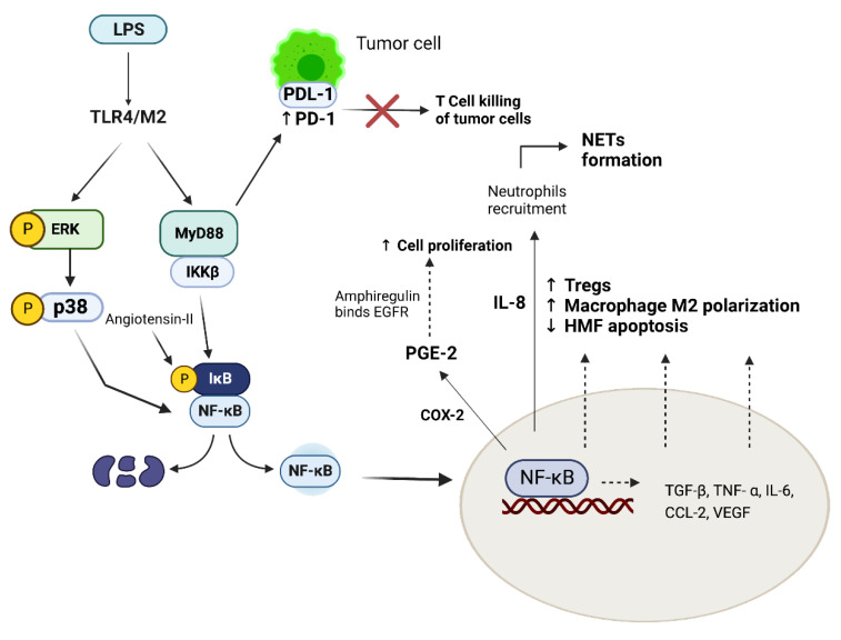
Figure 2.
Molecular pathways involved in tumor development and progression. LPS, through TLR-4, activates MyD88 and MAPK/ERK/p38 pathways, both capable of promoting NF-kB nuclear translocation—in particular, through MyD88 pathway, IKKB phosphorylates IkB, which detaches from NF-KB, allowing nuclear translocation. This mechanism is favored by autocrine Angiotensin-II. Once in the nucleus, NF-kB activation leads to the transcription of multiple cytokines. COX-2 promotes PGE2 production, which stimulates the release of amphiregulin, an EGFR ligand involved in cell proliferation. IL-8 is involved in neutrophils recruitment, with subsequent NETs formation. Moreover, NF-kB activation leads to a reduced apoptosis of HMF and HSC, increases the number of Tregs and enhances M2 macrophages polarization, altering the immune tumor microenvironment, thereby promoting tumor cells immune escape. LPS/TLR-4/MD2 pathway also increases PD-1/PDL-1 bond, blocking T cell killing of tumor cells. Abbreviations: LPS: lipopolysaccharide; TLR-4: toll-like receptor 4; MAPK: mitogen-activated protein kinase; IKKB: inhibitor of nuclear factor-κB (IκB) kinase (IKK) complex; NF-kB: nuclear factor NF-kappa-B; COX-2: cyclooxygenase-2; PGE2: E2 prostaglandin; EGFR: Epidermal growth factor receptor; IL-8: Interleukin-8; NETs: neutrophil extracellular traps; HMF: hepatic myofibroblasts; HSC: hepatic stellate cells; TGF-β: tumor growth factor- β; TNF-α: tumor necrosis factor α; IL-6: interleukin 6; CCl-2: chemokine (C-C motif) ligand 2; VEGF: vascular endothelial growth factor; PD-1: programmed death-1; PDL-1: programmed death ligand-1; Tregs: regulatory T cells. Created with BioRender.com.

