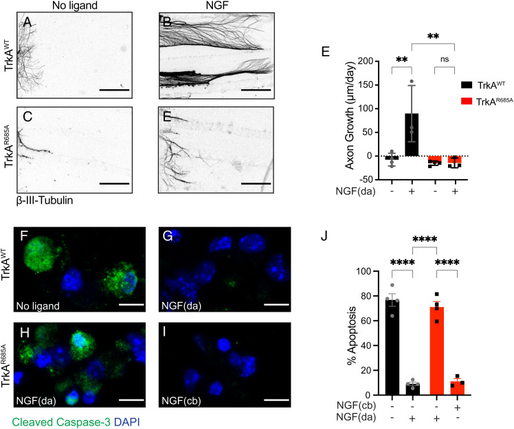
Fig. 4.
Impaired NGF-dependent axon growth and neuron survival in TrkAR685A neurons. (A–D) NGF-dependent axon growth is abolished in TrkAR685A neurons. Compartmentalized cultures of sympathetic neurons from P0-P3 TrkAR685A or TrkAWT mice were either deprived of NGF by including anti-NGF in media bathing both cell body and axon compartments (A and C) or maintained with NGF (30 ng/mL) added solely to distal axon compartments (B and D). The caspase inhibitor, BAF (50 μm), was included in all analyses to prevent cell death. Panels are representative images of axons immunostained with anti-β-III-tubulin at the end of the analyses. (Scale bar, 360 μm.) (E) Quantification of rate of axon growth (μm/d assessed in 24-h intervals for a total of 72 h). Results are means ± SEM from three to four independent experiments. At least 10 to 15 axons per condition per experiment were analyzed. **P < 0.01, two-way ANOVA and Tukey–Kramer post hoc test. (F and G) NGF (30 ng/mL) added only to distal axons (da) promotes survival of TrkAWT neurons compared to the no ligand condition, as assessed by cleaved caspase-3 immunostaining. (H and I) Neuronal survival is compromised in TrkAR685A neurons when NGF (30 ng/mL) is present on distal axons (da) but not when NGF is added directly to cell bodies (cb). Neuronal nuclei were labeled with DAPI (blue). (Scale bar, 5 μm.) (J) Quantification of neuronal apoptosis by determining the percentage of neurons that were cleaved caspase-3 positive. Only neurons that had extended axons into side chambers, defined by the retrograde accumulation of fluorescent microspheres, were scored for apoptosis. Results are means ± SEM from three independent experiments. At least 20 neurons per condition were assessed per experiment. Two-sample t test with multiple comparisons and Tukey–Kramer post hoc test, ****P < 0.0001.

