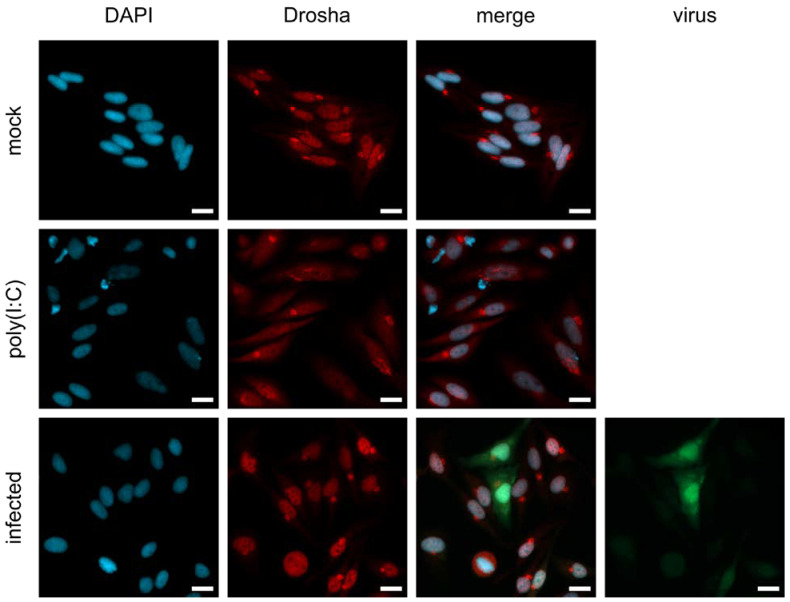
Figure 4.
Localization of Drosha upon MeV infection. Nuclear localization experiments with immunofluorescence staining of Drosha. HeLa cells were mock-treated, and treated with 10 μg/mL poly(I:C) or infected with MeV ld-EGFP at MOI 0.3 (green). Drosha (red) and the nucleus (blue) were stained with antibody and DAPI, respectively. Bright spots in the Drosha staining are considered staining artifacts. Scale bar: 20 µm.

