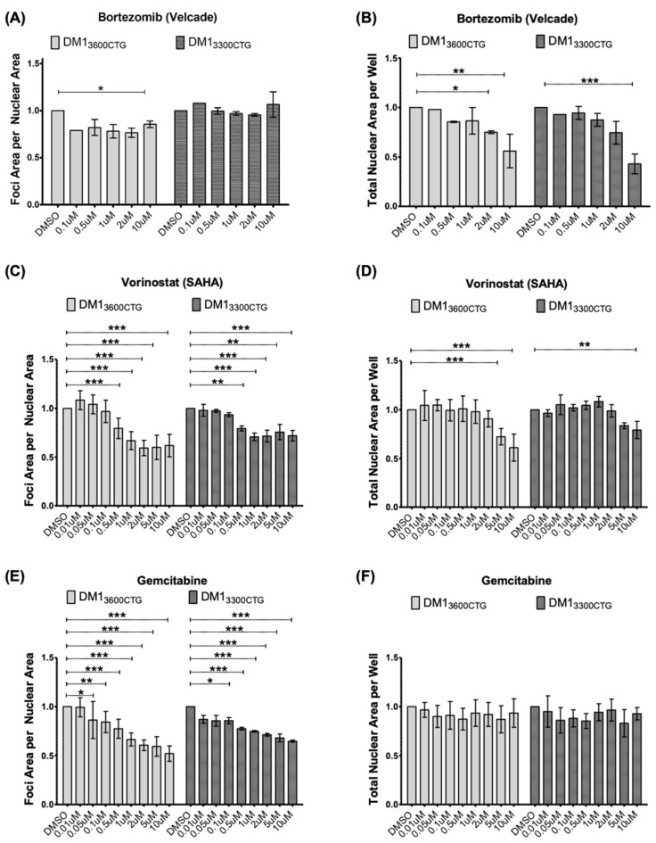
Figure 2.
Bortezomib minimally reduced foci; vorinostat (SAHA) and gemcitabine reduced foci in both DM13600CTG and DM13300CTG differentiated myoblasts. DM13600CTG and DM13300CTG myoblasts were serum-starved in 384-well plates for 7 days and treated with 0.1–10 μM of (A,B) bortezomib, (C,D) vorinostat (SAHA), and (E,F) gemcitabine. Post-treatment, cells were fixed with 4% PFA, DNA was stained with Hoechst and CUG RNA foci were probed by Alexa555-(CAG)10 fluorescent oligo. (A,C,E) Foci area per nuclear area and (B,D,F) total nuclear area per well (to assess treatment-associated toxicity) were quantified using Columbus and normalized to DMSO control; data is presented as fold-change relative to DMSO treatment (n ranges from 1 (for Bortezomib 0.1 µM) to 8, two-way ANOVA; error bars represent SD). * p < 0.05, ** p < 0.01, *** p < 0.001.

