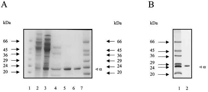
FIG. 1.
Purification of PGI from P. furiosus (A) and of recombinant PGI from transformed E. coli (B) as analyzed by SDS-PAGE. Protein was denatured in SDS and separated in 14% (A) or 12% (B) slab gels (8 by 7 cm) (23), which were stained with Coomassie brilliant blue R 250. (A) Lanes 1 and 7, molecular mass standards (Sigma), in kilodaltons; lanes 2 to 6, analysis of PGI after various steps of the purification procedure (lane 2, 100,000 × g supernatant; lane 3, Q Sepharose; lane 4, phenyl-Sepharose; lane 5, Superdex 200; lane 6, Uno Q1). (B) Lane 1, molecular mass standards (Sigma); lane 2, purified recombinant PGI.

