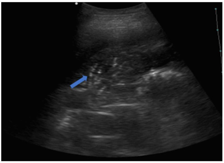
Figure 10.
The image shows a complete lobar consolidation with air bronchograms (blue arrow) and minimal parapneumonic pleural effusion (lobar pneumonia). The air interface is completely lost, and the lung lobe is represented as a parenchymatous organ similar to the hepatic parenchyma (lung hepatization).

