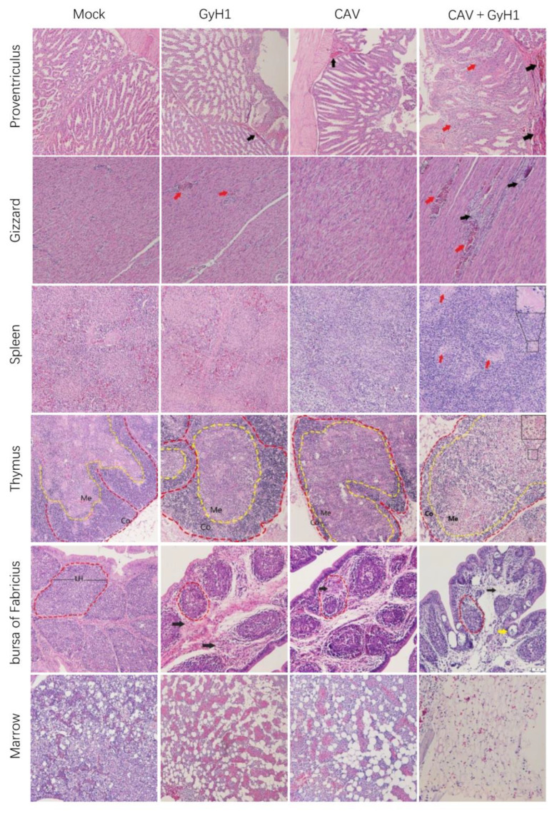
Figure 6.
Histopathological examination of immune and digestive organs (HE staining, 20×). Mucosal lamina propria exudation of erythrocytes in the proventriculus (black arrows), and structural disturbances resulting from necrosis of glandular tubule cells (red arrows); muscular hemorrhage (red arrows) and myofiber necrosis (black arrows) in the gizzard; splenic lymphocytes were lost, and germinal centers were enlarged (red arrows); decreased thymic cortical area (Co) and enlarged medullary substance region (Me); bursal lymphoid follicles (LH) are miniaturized with connective tissue hyperplasia (black arrow) and lymphatic follicular emptying (yellow arrow); adipocytes replace myeloid cells in the bone marrow.

