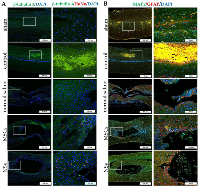
Figure 11.
IF staining of SCI tissue 8 weeks after transplantation by β-tubulin 3, HuNu, MAP2, and GFAP. (A) Images of IF staining by β-tubulin 3 (green) co-stained with HuNu (red), scale bars = 500 µm (left) and 100 µm (right). (B) Images of IF staining by MAP2 (green) co-stained with GFAP (red), scale bars = 500 µm (left) and 100 µm (right).

