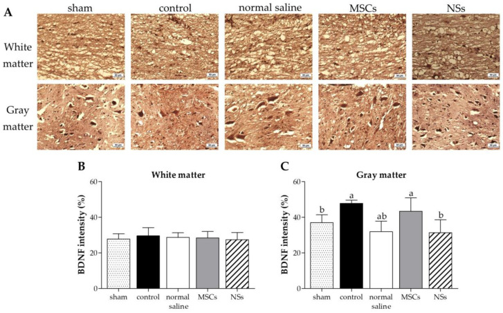
Figure 14.
Immunohistochemistry (IHC) staining of SCI tissue at 8 weeks after transplantation. (A) IHC staining images of BDNF at white matter and gray matter; brown particles were converged around the cavity of the spinal cord, scale bar = 50 µm. BDNF intensity results at (B) white matter and (C) gray matter of SCI tissue. Data were shown as mean ± S.D. with different lower-case letters, and are significantly different at p < 0.05.

