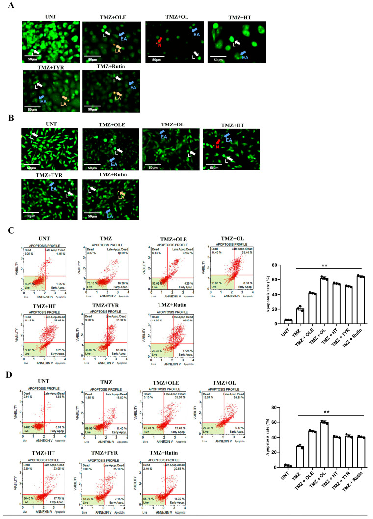
Figure 12.
Viability of human GB cells after treatment with TMZ + OLE, TMZ + OL, TMZ + HT, TMZ + TYR, and TMZ + rutin. AO/PI staining of T98G (A) and A172 cells (B). The green cells with a granular nucleus located on one side indicate apoptosis. In contrast, a circular nucleus uniformly distributed in the center of the cell indicate a cell in interphase. The red cells with an inapparent outline indicate necrosis, dissolved or near disintegration. The color-coded arrows indicate the following: alive cells in white, early apoptosis in blue, late apoptosis in orange, and necrosis in red. (C) TMZ-only, TMZ + OLE, and TMZ + OLE phenolics induced apoptosis in T98G and (D) A172 cells. The adjusted p-values were calculated using one-way ANOVA and Tukey’s post hoc tests. ** p < 0.0001 compared to untreated cells; n = 3. UNT: untreated, TMZ: temozolomide, OLE: Olea europaea leaf extract, OL: oleuropein, HT: hydroxytyrosol, TYR: tyrosol. L: alive cells, EA: early apoptosis, LA: late apoptosis, N: necrosis.

