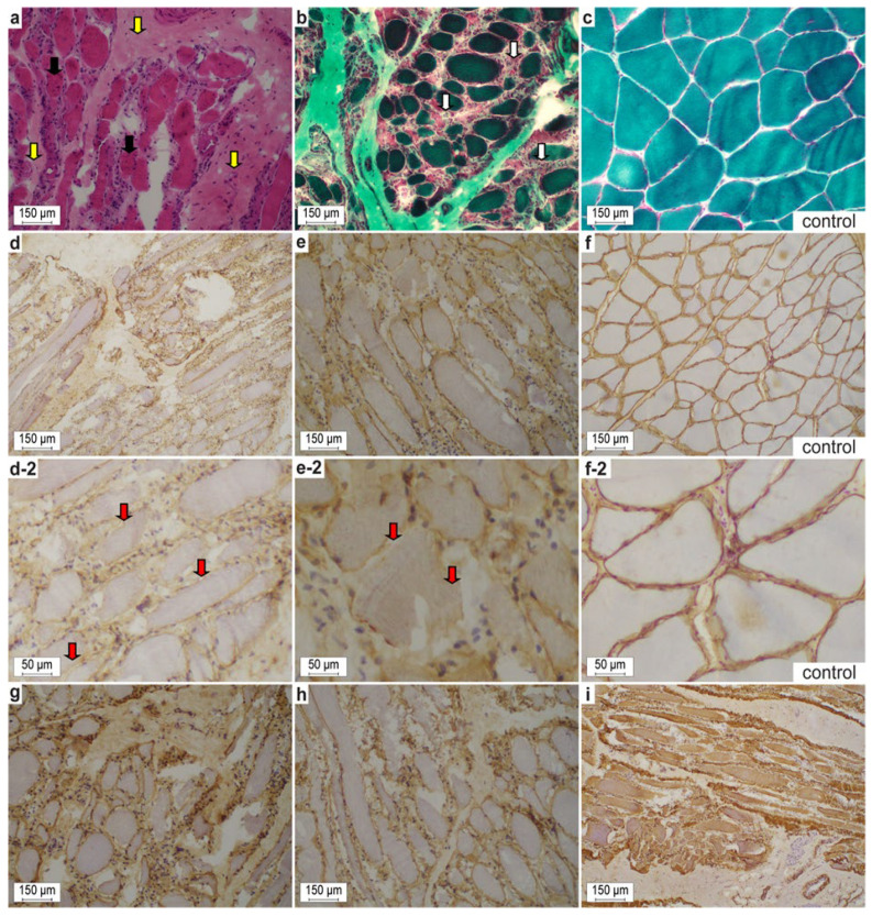
Figure 2.
(a,b,d,e,g–i) Representative cryosections of the affected gastrocnemius muscle from the dystrophic Maine Coon crossbred domestic cat #1. (a,b) Noteworthy are the variability of myofiber sizes, degenerative (groups of necrotic fibers with myophagocytosis, white arrows) and regenerative muscle features (small fibers with prominent vesicular central nuclei; multiple nuclear internalization, black arrows), and endomysial fibrosis (yellow arrows) in affected cat #1. (d,e) On immunohistochemistry, membranous dystrophin often is discontinuous or even absent in between individual fibers (red arrows). Moreover, γ- and β-sarcoglycans (g,h) and desmin (i) were diminished and interrupted. (c,f) control sections from a European domestic shorthair cat. (a) H&E stain. (b,c) Gomori trichrome stain according to Engel. (d,f) Dystrophin 1 (1:100, DYS1). (e) Dystrophin 2 (1:100, DYS2) at regular and increased magnification. (g) γ-sarcoglycan (1:100, γ-SARC). (h) β-sarcoglycan (1:50, β-SARC). (i) desmin (1:50, Desmin Clone D33 M0760). (c,f) Control sections of a domestic short hair cat.

