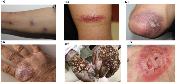
Figure 4.
Atypical morphology of CL lesions: (a) sporotrichoid-type lesions on the upper extremity of the CL patient; (b) lupoid form of lesions observed in arm of the CL patient; (c) psorasiform type of CL lesion on the elbow of the patient’s right arm; (d) eczematoid-type CL lesion on back of the hand; (e) verrucous-type CL lesion on back of the hand; 4 (a–e). [158]; and (f) multiple erythematous lesions with indurated margin and necrotic-purulent base mimicking pyoderma gangrenosum [159].

