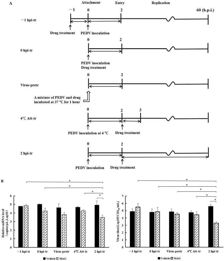
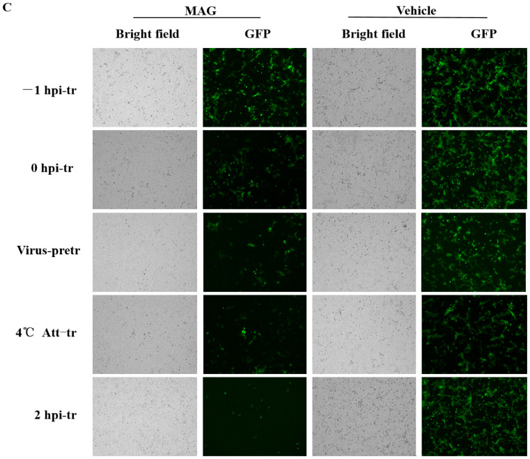
Figure 3.
rPEDV-∆ORF3-GFP titers, M protein mRNA levels and GFP expression images after MAG treatment at different stages of the virus life cycle. (A) Chronology of MAG treatments and assay operations. For the sake of clarity, only the attachment, entry and replication stages of the virus life cycle are shown. In −1 hpi-tr samples, cells were treated with MAG 1 h before infection with virus. In the 0 hpi-tr samples, MAG was applied to the cells at the same time as the virus. In virus-pretr samples, the reaction components were first incubated at 37 °C for 1 h before adding to the cells. In 4 °C Att-tr samples, viruses were first incubated with Vero cells at 4 °C for 2 h. The inoculum was then removed and the temperature was raised to 37 °C prior to drug treatments and further incubation. In 2 hpi-tr samples, MAG was applied 2 h after virus infection. (B) 60 hpi titrations and expression of rPEDV-∆ORF3-GFP M protein mRNA using different treatment protocols. Data are presented as the mean ± SEM of the three independent experiments (* p < 0.05). (C) GFP expression images of rPEDV-∆ORF3-GFP using different treatment protocols as indicated.


