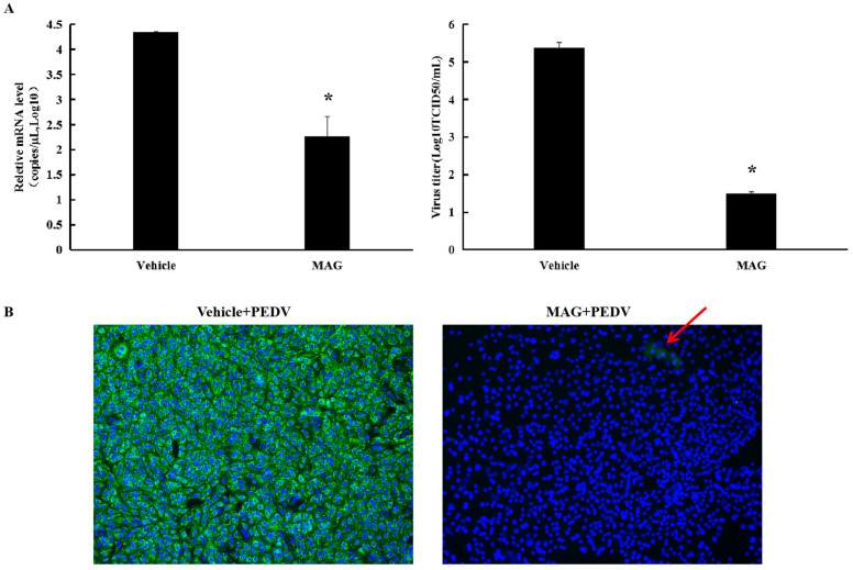
Figure 4.
MAG inhibition of PEDV replication in vitro. (A) Vero cells were infected with PEDV for 2 h, after which the inoculum was removed and fresh maintenance medium containing 30 μM MAG was added. Supernatants were collected 36 hpi, and virus titers (TCID50) and PEDV mRNA levels were determined. Vehicle controls consisted of DMSO at a final concentration of 0.015%. Data are presented as the mean ± SEM of the three independent experiments (* p ˂ 0.05). (B) Vero cells were treated with 30 μM MAG for 1 h and then infected with PEDV DR13att. The inoculum was removed after 2 h incubation and fresh maintenance medium containing 30 μM MAG was added. Vero cells were fixed and subjected to immunofluorescence staining using anti-PEDV M polyclonal antibody 24 hpi. Red arrow indicates the rear immunofluorescence staining of PEDV M protein (green) in MAG+PEDV treated cells.

