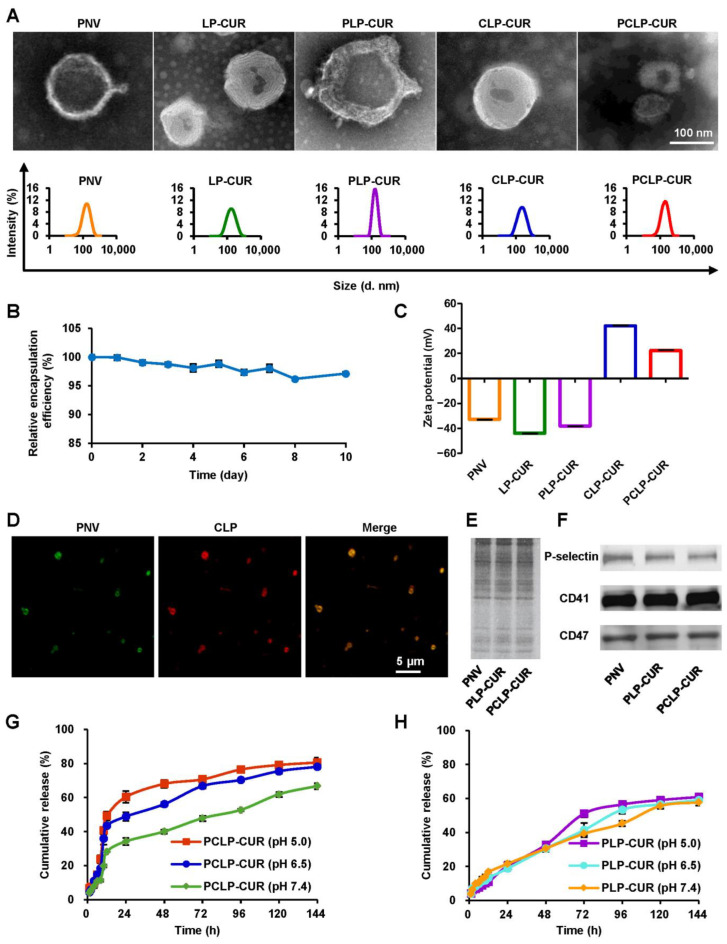
Figure 1.
Characterization of PCLP-CUR. (A) Morphologies observed by transmission electron microscopy (upper panel) and size distributions observed by a Zetasizer (bottom panel). (B) Stability of PCLP-CUR at 4 °C for 10 days in terms of relative encapsulation efficiency. (C) Zeta potentials of various nanoparticles observed by a Zetasizer. (D) Fluorescence images of PCLP observed by confocal laser scanning microscopy. The PNV and CLP were labeled with DiO (green) and DiD (red), respectively. Yellow color indicated a colocalization between the two corresponding signal. (E) The results of SDS-PAGE analysis. (F) Western blot analysis of key platelet membrane proteins. (G) Drug release characteristics of PCLP-CUR at pH 5.0, 6.5 or 7.4. (H) Drug release characteristics of PLP-CUR at pH 5.0, 6.5 or 7.4.

