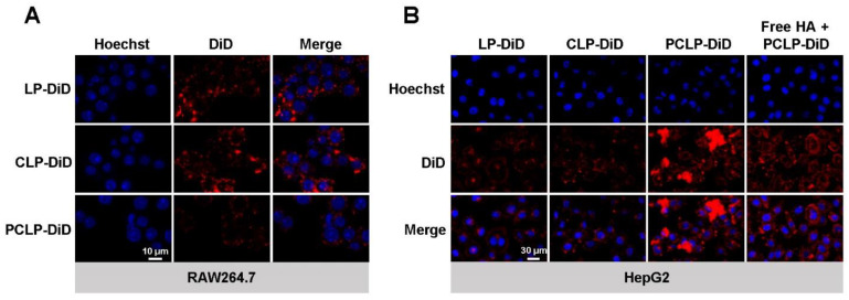
Figure 2.
Efficient in vitro delivery of PCLP observed by confocal laser scanning microscopy. (A) Fluorescence images of RAW264.7 cells incubated with different liposomes for 2 h. (B) Fluorescence images of HepG2 cells incubated with different liposomes for 2 h after a 3 h preincubation with or without free HA. Nuclei were labeled with Hoechst 33342 (blue). All nanoparticles were labeled with DiD (red).

