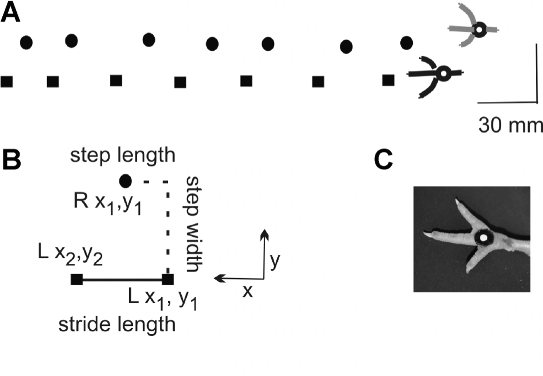
FIGURE 1.

Kinematic methods. (A) Left (square) and right (circle) foot placements within the recording region are shown for one walk trial as the chick progressed from right to left. The scale bar of 30 mm applies to both x and y directions.(B) Methods for measuring spatial parameters are shown. Stride length was defined as the distance between consecutive foot strikes of the same foot (i.e., Lx2 − Lx1). Step length was equal to the distance between consecutive strikes of the left and right foot(i.e., Rx1 − Lx1). Step width was equal to the distance between consecutive left and right foot strikes in the mediolateral direction (i.e., Ry1 − Ly1). (C) Circular markers were applied to the metatarsal pads of both feet. A white dot and black contrasting surround optimized automatic tracking during digitizing of foot placements.
