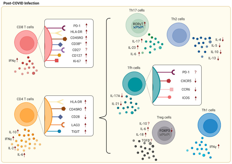
Figure 2.
Changes in surface markers and cytokines following SARS-CoV-2 infection. Increased expression of surface markers including PD-1, Ki-67, HLA-DR, CD45RO, CD38hi, and CD127 was found on CD8+ T cells, while CD27 shows a contradicting result. Upregulation of LAG-3, TIGIT, HLA-DR, and CD45RO as well as downregulation of CD28 were detected on CD4+ T cells. Included in CD4 T cell subsets are Th1, Th2, Th17, Tfh, and Treg cells. Increased populations of Th1 cells along with the production of IFNγ predominated following SARS-CoV-2 infection. Conversely, low numbers of Th2 cells along with abnormal secretions of associated cytokines were found in mild patients. As for Tfh cells, lower expressions of CXCR5 and CCR6 and higher expression of ICOS were observed, while PD-1 expression had conflicting results. There were contradicting studies for Treg cells with some suggesting that high populations of Treg cells were associated with increased secretion of IL-10, TGF-β, IL-6, and IL-18. On the other hand, some researchers indicated a reduction in Treg cells, with lower expression of FOXP3, TGF-β, and IL-10. Lastly, an increased percentage of Th17 cells with high expression levels of RORγt and increased secretion of signature cytokines are found post-SARS-CoV-2 infection.

