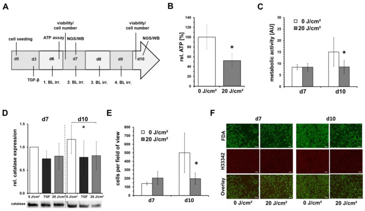
Figure 2.
Low-dose blue light irradiations inhibit fibroblast proliferation. Shown are mean ± SD values of 5 independent experiments, * p < 0.05 as compared to the control values. Human dermal fibroblasts were irradiated with low, sub-toxic doses (20 J/cm2) of blue light (420 nm). (A) Flowchart of experimental procedure. Human dermal fibroblasts were seeded on day 0 for experiments and grown for 6 days. On d6–d9, fibroblast cultures were irradiated by blue light (BL; 420 nm; 33.6 mW/cm2) and cell culture media was changed on a daily basis. Determinations of cell viability, cell number, intracellular ATP concentration, sample preparation for next generation sequencing (NGS) and Western blot (WB) were performed as indicated. As positive control, TGF-β (10 ng/mL) was added for some experiments. (B) Results of intracellular ATP-concentration 1 h after irradiation on day 6. (C) Metabolic activity assessed by a resazurin-based assay and (D) relative catalase protein expression assessed by western blot 16 h or 24 h, respectively, after one irradiation on day 7 and four irradiations on day 10. (E) Quantitative determination of dead/live cell ratios obtained from live cell imaging using Hoechst 33342- and fluorescein diacetate (FDA) staining. (F) Shown are representative microphotographs of Hoechst 33342- and FDA-stained fibroblast cultures (50×). Viable cells show a green fluorescence signal of cytoplasma by FDA. Nuclei were stained by Hoechst 33342, and the signals were colored in red for better visualization. Bars = 200 µm.

