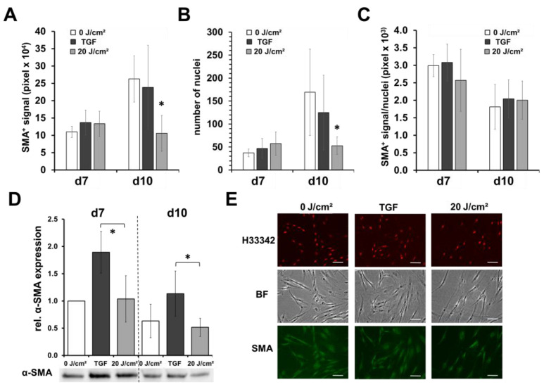
Figure 3.
Blue light does not induce myofibroblast differentiation. Shown are the means ± SD of 5 experiments with different patients (* p < 0.05). Human dermal fibroblasts were irradiated with low, sub-toxic doses (20 J/cm2) of blue light (420 nm). On day 7 (24 h after 1st blue light irradiation) and on day 10 (4× blue light irradiations), fibroblasts were fixed, subsequently immunocytochemically stained with antibody against α-smooth muscle actin (α-SMA), a myofibroblast marker, and evaluated by fluorescence microscopy. As positive control, TGF-β (10 ng/mL; TGF) was added from d3. (A) Determination of the pixel number of the positive α-SMA fluorescence signal or (B) number of nuclei (Hoechst 33342+) in a field of view at 100× magnification (4 fields of view/well). (C) Ratio of α-SMA/number of nuclei. (D) Relative protein expression of α-SMA assessed by Western blot analysis. (E) Shown are representative microphotographs (200×) of α-SMA- and Hoechst 33342-stained fibroblast cultures on day 10 (bright field; BF). Hoechst 3342 signals were colored in red for better visualization. White bars = 50 µm.

