
Figure 4.
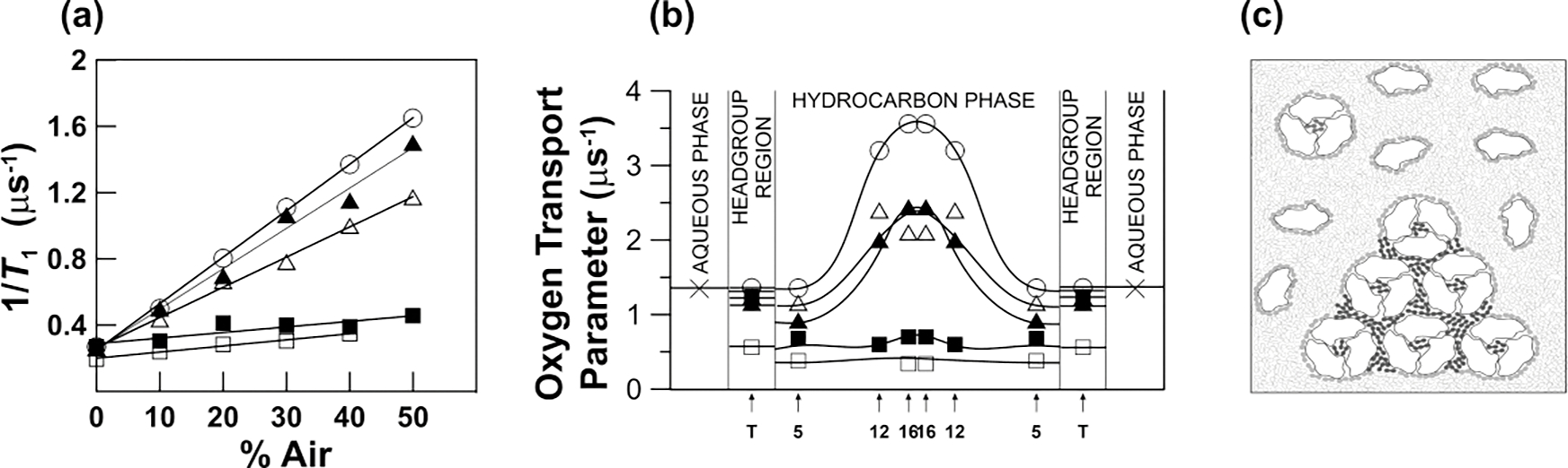
Steps used in the discrimination of membrane domains using the DOT method. (a) Spin lattice relaxation rates of 1-palmitoyl-2-(12-doxylstearoyl)phosphatidylcholine (12-PC) plotted as a percentile of air in the gas mixture equilibrating the membrane suspension at 30°C. Symbols are for the DMPC bilayer without BR (○), with BR/DMPC = 1/80 (Δ), with BR/DMPC = 1/40 (▲,■), and for purple membranes isolated from Halobacterium halobium (□). values were extrapolated to 100% air and OTP was calculated for each domain. As indicated in Eq. 1, the SR signals obtained for the DMPC bilayer without BR, with BR/DMPC = 1/80, and for purple membranes were successfully fitted to single exponentials, giving single values of the OTP for all spin labels (using Eq. 1). SR signals obtained for the DMPC bilayer with BR/DMPC = 1/40 were successfully fitted only to double exponential functions, giving two values of the OTP for each spin label (using Eqs. 4 and 5). (b) Profiles of the OTP values obtained at 30°C from different PL spin labels across the DMPC bilayer without BR (○), with BR/DMPC = 1/80 (Δ), with BR/DMPC = 1/40 (▲,■), and across purple membranes (□). When BR is in the monomeric form, only one bulk-plus-boundary lipid domain is present (Δ). When BR is aggregated, two lipid domains coexist: bulk-plus-boundary domain (▲) and trapped lipid domain (■). Arrows indicate approximate locations of nitroxide moieties of n-PCs and n-SASLs used in these investigations. T indicates T-PC. The symbol × indicates OTP in the aqueous phase. (c) Schematic drawing of the lateral organization of bacteriorhodopsin and lipid molecules in the reconstituted membrane of BR and DMPC at a BR/lipid ratio of 1/40. Phospholipid molecules are indicated as an open and closed figure-eight-shaped phospholipid cross section. Phospholipids in the bulk domain are open, in the boundary are grey, and in the SLOT domain (trapped-lipid domain) are dark. Lipids in the SLOT domain are trapped between trimers and oligomers of trimers of the BR. The schematic shapes of molecules are drawn on the base of the electron microscopy studies [54]. Data for Fig. 4a and Fig. 4b are reproduced with permission from Ref. [55]. Copyright 2022, American Chemical Society.