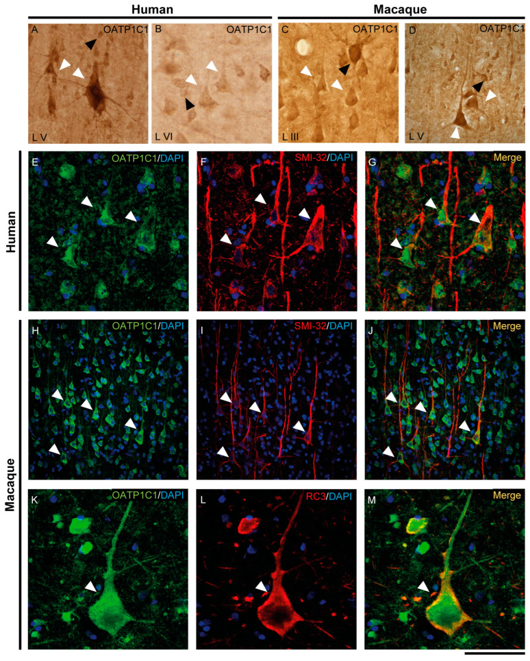
Figure 4.
Expression of OATP1C1 in pyramidal neurons of the human and macaque motor cortex. Representative brightfield photomicrographs show immunostaining for OATP1C1 in layers V (A) and VI (B) of the human motor cortex and layers III (C) and V (D) of the macaque motor cortex. (E–G) Confocal microscope images for OATP1C1 (green, E) and the pyramidal neuron marker SMI-32 (red, F) immunostaining in the human cortex. (G) Merged image showing the colocalization of both signals. (H–M) Confocal microscope images for OATP1C1 (green, H,K), SMI-32 (red, I) and neurogranin/RC3 (red, L) immunostaining in the macaque cortex. (J,M) Merged confocal photos, respectively, showing the colocalization of the signals. Counterstaining with DAPI (blue) shows nuclei of all cells. White arrowheads point to pyramidal neurons. Note that the signal can be identified in the membrane, cytoplasm, and apical and basal dendrites. Black arrowheads point to neurons with interneuronal morphology. L III–VI: layers of the cerebral cortex. Scale bar = 120 μm (A,D,H–J), 60 μm (B,C), 40 μm (E–G), 50 μm (K–M).

