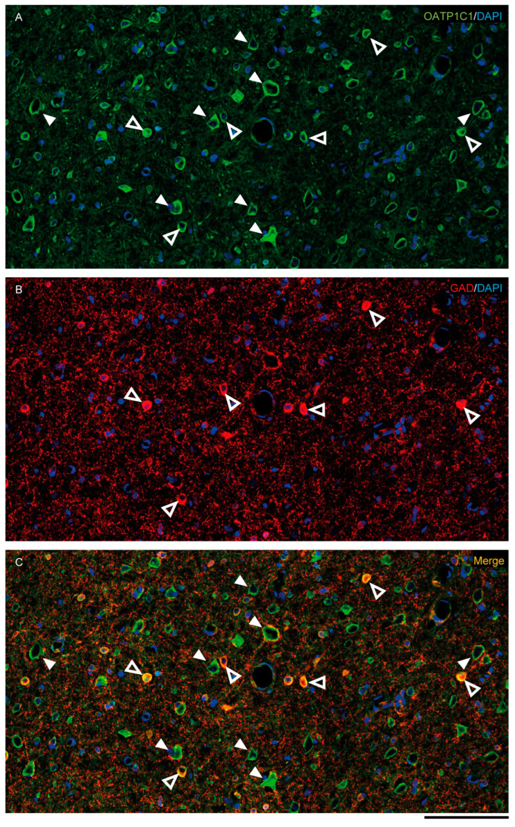
Figure 5.
Expression of OATP1C1 in the macaque cortical GABAergic neurons. Confocal tile scan microscope images of a macaque cortical section double-stained for OATP1C1 (green, A) and the interneuronal marker GAD (red, B). (C) Merged image showing the colocalization of both markers only in small cells surrounding pyramidal neurons. Counterstaining with DAPI (blue) shows nuclei of all cells. White arrowheads point to pyramidal neurons. Hollow white arrowheads point to double-labeled interneurons. Note that OATP1C1 is expressed in practically all GAD-immunopositive neurons. GAD: glutamic acid decarboxylase. Scale bar = 100 μm.

