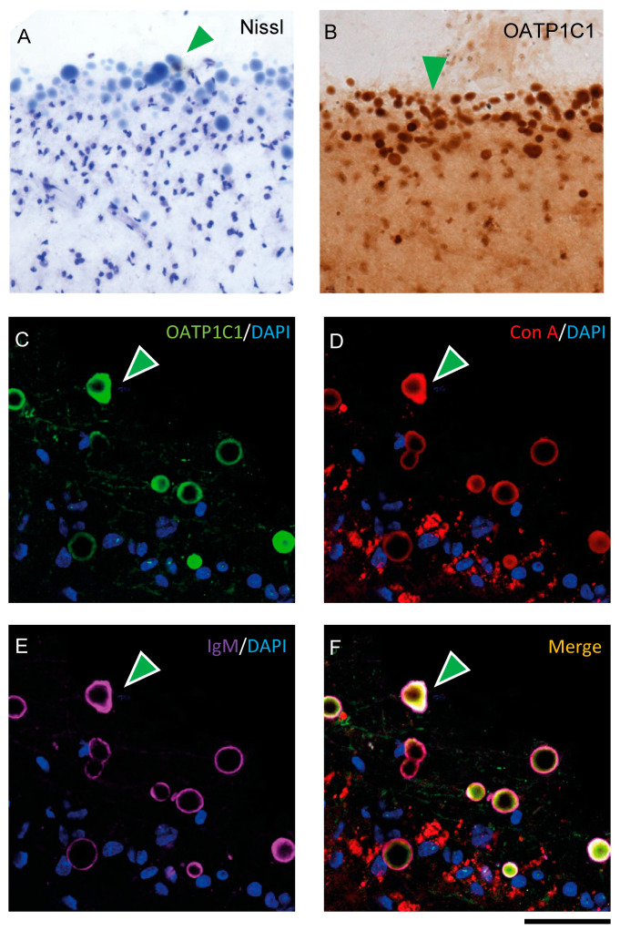
Figure 8.
Expression of OATP1C1 in Corpora amylacea of the human motor cortex. (A,B) Representative brightfield photomicrographs of Nissl-stained (A) and OATP1C1 immunostained (B) sections in which green arrowheads point to numerous spherical vesicles located in the subpial region and layer I of the human motor cortex. (C–F) Confocal microscope images of triple labeling for OATP1C1 (green, C), and the two Corpora amylacea markers Con A (red, D) and IgM (purple, E). The merged image (F) shows the total colocalization of the three markers (green arrowheads). Counterstaining with DAPI (blue) shows nuclei of all cells. Note that the vesicles do not contain a blue nucleus. Con A: Concanavalin A. Scale bar = 50 μm (A,B), 40 μm (C–F).

