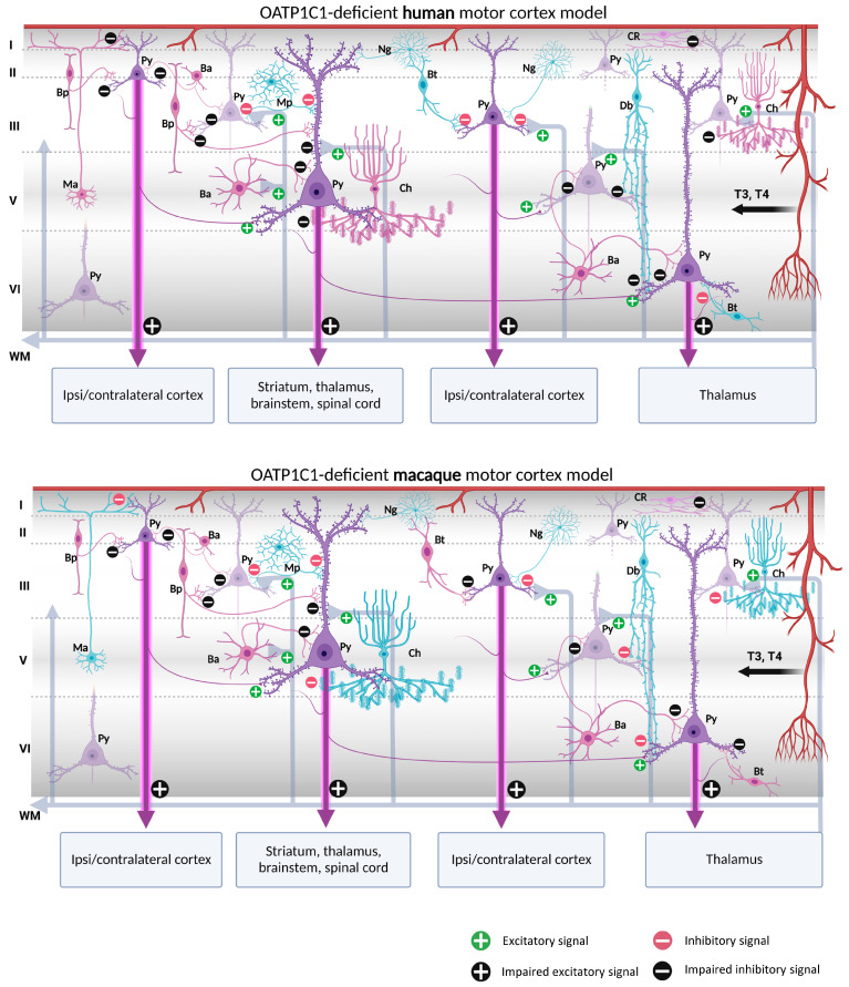
Figure 11.
Models of human and macaque OATP1C1-deficient motor cortices. The models include our results and show possible cortical functional microcircuitry physiopathogenic mechanisms. The models might explain some of the symptoms generated in OATP1C1-deficient syndromes. Pyramidal cells and interneurons expressing OATP1C1 are shown in purple. Interneurons non-expressing OATP1C1 are shown in blue. The lack of T4 (and thus decreased deiodination by the astrocytes) may cause trophic deficits in pyramidal cells and interneurons, generating hyperexcitation on pyramidal cells due to the possible abnormal inhibitory function of the interneurons. Abnormalities in pyramidal cells may cause irregularities in their excitatory output signaling to intrinsic and extrinsic targets, producing symptoms such as spasticity, hypertonia, hyperreflexia, epilepsy and others, depending, for example, on the final dendritic activity. Ba: basket cell, Bp: bipolar cell, Bt: bitufted cell, Ch: chandelier cell, CR, Cajal–Retzius cell, Db: double bouquet cell, Mp: multipolar cell, Ma: Martinotti cell, Ng: neurogliaform cell, Py: pyramidal cell, L I–VI: layers of the cerebral cortex, WM: white matter. (Scheme based on Szentágothai, 1975 [139] and Jones, 1993 [140]; created with BioRender.com).

